

Bad Breaks
Though their hips were shattered, three people walk again—their fractures repaired through the most complex, technically demanding type of orthopaedic trauma surgery.

A postoperative image showing the hardware that secures the joint and allows it to heal properly.
![]()
It is critically important to get this reconstruction just right, ideally within two millimeters of perfection.
![]()
"I have crashed into a wall and lived. I can do anything now."
Darren Hill
![]()
Jay Smith was heading home from a St. Louis Rams football game when his tire caught the edge of the road, his car skidded down an embankment, and it struck a tree.
St. Louis police officer Darren Hill was driving at 60 miles an hour when he lost control of his patrol car and slammed into a retaining wall.
Washington University sophomore Emily Levin was in-line skating in Forest Park when she traveled down a steep hill, into a designated crosswalk, and was struck by a passing bus.
ALL THREE—Smith, Hill and Levin—suffered "high-energy trauma," the kind of injury that occurs in high-speed motor vehicle accidents and falls from more than 10 feet. They did not fit the usual demographic for such mishaps: young men from 18 to 45, driving recklessly and often drunk, late at night or on the weekend. But each of them emerged from his or her accident with the same, devastating hip injury, an acetabular fracture.
"Think of the patient’s hip as a ball and a socket," says Joseph A. Borrelli Jr., MD, assistant professor of orthopaedic surgery, who performed the surgery on each. "As a result of the high-energy trauma, the ball is driven into the socket, or ‘acetabulum,’ and it fractures. Since the socket is part of the pelvis, and in a very difficult spot to get to, the surgery for this injury is associated with many potential problems."
Luckily, acetabular fractures are not common; there are probably fewer than 10,000 in the United States each year. There are also fewer than a dozen orthopaedic surgeons nationwide who specialize in such complex reconstructions—and Borrelli is among this select group. With his partner, William M. Ricci, MD, he has made Barnes-Jewish Hospital (BJH) a regional referral center for these fractures, treating patients from as far away as Arkansas, Tennessee, Texas and South Dakota. He is also actively developing new clinical protocols and basic research projects related to these injuries.
In Jay Smith’s accident, his acetabulum splintered, but he had no other injuries, just severe pain in the area of his hip. That pain is also what Darren Hill remembers, along with the sight of his fractured left forearm, left and right tibias, and right ankle. Emily Levin was the most severely injured, with an acetabular fracture and an open pelvic fracture, visible through a large laceration. Drifting in and out of consciousness, she was rushed to the BJH emergency room, where the trauma team worked feverishly to save her life. Soon her father, a New York orthopaedic trauma surgeon, was making calls to colleagues asking them to recommend surgeons who could perform her acetabular surgery—and discovered that two specialists were faculty at Washington University.
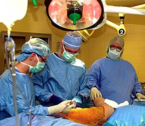
Joseph A. Borrelli Jr., in surgery with Charles Mettach, surgical technician (right) and third-year resident Gregory Della Rocca, MD (left).
Shortly after their accidents, all three patients underwent acetabular reconstruction, the most complicated, technically demanding kind of orthopaedic trauma surgery. To get to the patient’s shattered hip socket, the medical team—composed of surgeons, anesthesiologists, skilled nurses and X-ray technicians—has to skirt critical arteries, veins and nerves. Any mistake could cause serious bleeding or nerve injury that might mean permanent weakness or paralysis. When they reach the socket, surgeons must move in from behind and piece it together like a jigsaw puzzle. Then they fix it in place, screwing in plates up to eight inches long that remain in the body.
It is critically important to get this reconstruction just right, ideally within two millimeters of perfection. Any displacement in the articular surface—where the ball and socket meet and "articulate," or rub together—will expose the injured cartilage to stresses that will eventually lead to its deterioration. These displacements are referred to as "steps and gaps"—the bane of an orthopaedic trauma surgeon. And there are other potential complications as well. "You also want to be as sure as you can that your screws don’t enter the joint space, because that would be like trying to walk with a pebble in your shoe," says Borrelli.
Performing this type of surgery requires enormous stamina and concentration, since operations may be up to 12 hours long. One blessing is the timing. Unlike some surgical procedures that must be done immediately, acetabular fractures can often wait for several days, even a few weeks. "But you still have to enjoy being up when half the world is sleeping, doing long, challenging operations," says Borrelli, who did his orthopaedic trauma fellowship at Tampa General Hospital and joined the Washington University faculty five years ago.
In a procedure like this, so fraught with potential complications,
planning is crucial. But X-rays alone may not be enough since bones block
good views of the acetabulum; it is also hard to roll an injured, hurting
patient into position for the necessary images. So Borrelli has become
a proponent of adding CT scans to the planning mix. As part of his research,
he also has compared the use of X-rays and CT in assessing patients’
postoperative outcome. In one study, he looked at 15 patients to see how
many had step and gap problems: X-ray indicated a single deformity, while
CT was more accurate, showing that six had a significant displacement.
After his operation, Jay Smith spent seven days recovering at BJH. He recalls his first, tentative trip to the water cooler, bent over a walker. Because of his multiple injuries, Darren Hill had six surgeries, spending eight weeks in BJH followed by nearly four months in a nursing home. For two of those months he was bedridden, forbidden even to sit at a 90-degree angle; gradually, he began physical therapy and progressed from a walker to crutches to a cane. Emily Levin was unconscious for 12 days, then wide awake—sleepless—for a full week. In BJH for six weeks, with worried family and friends at her bedside night and day, she had nine surgeries, including two orthopaedic procedures. In early June, an air ambulance ferried her to a rehabilitation hospital near her home, where she began aggressive therapy.
No matter how hard surgeons work to eliminate steps and gaps, says Borrelli, some patients will go on to develop arthritis just because of the trauma to their cartilage. In his basic science research, Borrelli has studied this phenomenon—first by looking at the effect of high-impact injuries on bovine cartilage; and more recently, thanks to funding from the Orthopedic Research and Education Foundation, by developing an animal model for these cartilage injuries, the only such in vivo model in existence.
Already, he has found that there can be irreversible cartilage damage after trauma, even without an acetabular fracture. The reason may emerge in another study, funded by the Orthopaedic Trauma Association, in which he is looking closely at "apoptosis" or programmed cell death, a process that can be stimulated in cartilage by high-impact injury. Physicians have to find new ways to keep these cells alive, he says, if they want to reduce the incidence of post-traumatic arthritis.
During the postoperative recovery period—which often lasts nine months or more—Borrelli and his team carefully monitor each patient’s progress. Working with Jack R. Engsberg, PhD, of the Human Performance Laboratory, they use video cameras and other tools to measure stride length, walking speed and body angles, and compare that with data on people who have not been injured.
They also assess muscle strength and ask patients to complete
a detailed Musculoskeletal Functional Assessment questionnaire (MFA).
In studying the results, they have found that good outcome seems to correlate
with strength in particular muscles around the hip. Now they have applied
for an NIH grant to look at rehabbing those muscles in patients who scored
poorly on the MFA to see whether that improves their quality of life.
Three years out from his accident, Jay Smith says he is about 90 percent
back to normal. An insurance agent by day, he can still referee collegiate
sports by night, including some Washington University games. He is not
in pain, "but when it rains, or the weather changes, I notice it."
Nearly two years later, Darren Hill is hoping to be back on the police force full time in January; in fact, that goal was what kept him going from the start. "I have crashed into a wall and lived. I can do anything now," he says.
Astonishingly, Emily Levin took a full load of courses at SUNY– Stonybrook this past fall, just six months after her injury. She has some physical therapy ahead, as well as plastic reconstructive surgery. This January, she returns to Washington University and perhaps will even go skiing this winter.
"I can’t wait," she says. "That would be an understatement," adds her mother.
All say they are grateful to Borrelli and his staff, as well as the emergency room trauma teams that stabilize patients in the first critical hours after an accident.
"Having patients like Emily and Jay and Darren—seeing
their hips heal and watching them get on with their lives—that is
wonderful," says Borrelli. ![]()
![]()
![]()