
|
|
||
|
The New Microbiology Revolutionary technical advances deepen the understanding of fundamental life processes |
||||||
|
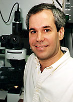
"It's not just streaking bacteria on plates anymore." Scott J. Hultgren, PhD |
MICROBIOLOGY ISN’T WHAT IT USED TO BE, and Scott J. Hultgren’s laboratory is a perfect example of how the field has changed. "It’s not just streaking bacteria on plates anymore," says Hultgren, PhD, Helen L. Stoever Professor of Molecular Microbiology. "The field has become a melting pot of disciplines working together to understand the molecular basis of infectious diseases." In the early days of microbiology, people studied the bacterium itself, simply trying to understand it. Next, investigators infected animals with a pathogen and studied the outcome. They also learned to isolate toxins and other products of microbes and then tested their effects on cells, cell components and animals. A long period followed when it was difficult to get beyond this descriptive science. The development of recombinant DNA technology broke that logjam and has had a profound effect on microbiological research—and on almost every field of medicine—that continues to the present day. Today, the dark world of microbial parasites is being illuminated by scientists who can eliminate, replace and monitor specific genes. Other methods enable researchers to tag and locate specific proteins within pathogens and to identify genes that are active and inactive in both parasite and host cells. The work is yielding valuable new insights into the relationship between the human cell and the infectious organism, into the balance between host and pathogen. |
|||||
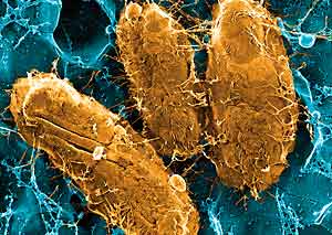
| Scott
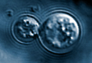
J. Hultgren, PhD A cross section of basic research is represented in the Hultgren laboratory’s study of the bacterium E. coli (right).
|
A multidisciplinary approach Hultgren’s research focuses largely on understanding the cause of urinary tract infections (UTIs) and developing more effective methods of treatment. Researchers in his laboratory work with investigators from other specialties to approach these questions from many angles. UTIs are caused by strains of Escherichia coli bacteria, occur predominantly in women, and frequently recur. Doctors have assumed that recurrent UTIs were caused by the repeated introduction of E. coli into the urinary tract, during sex or due to poor hygiene. Although the likely source of the originating infection, Hultgren and colleagues have discovered that the bacteria can latently infect bladder cells; activation can cause recurrence. Infecting E. coli bind to bladder cells using hair-like pili that contain a type of protein known as an adhesin. The adhesin binds with a receptor on the cell’s outer layer, triggering it to reach out and envelop the bacterium in a vacuole, or small pocket, within the cell. Hultgren’s laboratory is working to understand this process and to develop a vaccine that will block it. Other approaches his group is taking include crystallographers studying the structure of the adhesin-receptor interaction; immunologists examining the host’s immune response to the bacterium; cell biologists studying the pathways used by the invading bacteria to enter the cell; biochemists studying how the pili and adhesin proteins are formed, and geneticists investigating the genetic basis for the bacterium’s virulence. |
|||||
|
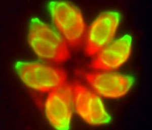
L David Sibley , PhD
|
High-resolution microscopy and genome sequencing L. David Sibley, PhD, associate professor of molecular microbiology, uses the latest tools of molecular science to learn how the parasite Toxoplasma gondii infects cells. Thought to infect 50 percent of the world’s population, Toxoplasma is a highly successful parasite. It causes toxoplasmosis, which often remains asymptomatic, but can result in encephalitis (a swelling of the brain) and sometimes in eye and lung disease. It is also an important cause of congenital infection that can result in severe birth defects. People acquire Toxoplasma by eating undercooked meat or by ingesting soil containing parasite spores. Enzymes in the intestine liberate the tiny, slipper-shaped parasites, which cross the intestinal lining and enter nucleated cells. There, each one forms a cyst and multiplies until the cell bursts, liberating about 250 parasites, each of which then crawls off to infect other cells. Sibley is working to understand the parasite’s unusual motility and how it recognizes and invades cells. Toxoplasma infects cells by striking them head-on, pushing in the membrane—like pushing in the side of a balloon with a finger—until it creates a small oval compartment in the cell. "It represents a very clever stealth mechanism," he says. "The host cell doesn’t know it’s been infected." Sibley and his colleagues use high-resolution confocal microscopy to examine how the parasite interacts with the host cell. A kind of light microscope that enables researchers to view a very thin slice of a cell, the technique produces a clearer image and greater resolution than the typical light microscope. Investigators are using confocal microscopy with living cells, as well as electron microscopy with fixed cells, to localize specific proteins in the parasite and in host cells. In collaboration with the School of Medicine’s Genome Sequencing Center, Sibley’s laboratory has sequenced regions of Toxoplasma genes and genes of related parasites that are active during different stages of infection. "This work reveals the inner workings of Toxoplasma, and it enables us to identify genes that are present in a wide range of organisms—including their human or animal hosts—and those that are unique to the parasite," says Sibley. "The latter group provides potential targets for vaccine development or for new drugs designed to combat infection." |
|||||
|
Tamara L. Doering, MD, PhD
|
RNA interference Tamara L. Doering, MD, PhD, assistant professor of molecular microbiology, studies Cryptococcus neoformans, a yeast-like fungus that causes cryptococcal meningitis. The disease strikes people with severely suppressed immunity and can be fatal if left untreated. Cryptococcus is a sugar-coated parasite with an outer capsule of carbohydrates that is essential for the organism to cause disease. Once inside the host, the organism churns out huge quantities of capsule material that accumulate in tissues and disrupt the body’s immune response. "We want to learn how the capsule is produced,"
says Doering, "and then we’ll try to develop compounds that
inhibit the process." Certain biological idiosyncrasies of Cryptococcus make such work more difficult than for some other organisms. Doering has shortcut some of the difficulties because the entire genome for Cryptococcus has been sequenced, and the data are available on-line. This information has enabled her to quickly identify genes that are likely to be important for production of the capsule. For example, transporter proteins move sugars from one area of the cell to another and are essential for construction of the capsule. By comparing bits of similar proteins found in other organisms with the DNA sequence for Cryptococcus, she can identify genes that seem likely to code for a transporter protein in Cryptococcus. But how does Doering know that her gene codes for a transporter protein? One way is to stop the gene from working to see how it affects capsule production. Doering does this using a technique developed only in the last few years, double-stranded RNA interference. When Cryptococcus makes a transporter protein, a gene is turned on in the cell nucleus, and a copy of the gene is made in the form of messenger RNA (mRNA). The mRNA, which consists of a single strand of RNA, then moves to the cytoplasm where the protein is assembled. Scientists have learned, however, that if even a short piece of double-stranded RNA is present that matches the structure of an mRNA, the latter is destroyed. This prevents the protein from being produced. "Once we understand the biology," says Doering, "we can also look for ways to outsmart the pathogen." |
|||||
|
William E. Goldman, PhD
|
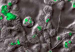
Assembling microarrays William E. Goldman, PhD, professor of molecular microbiology, studies Histoplasma capsulatum, the parasitic fungus that causes histoplasmosis. This fairly common infection is usually brought under control by a healthy immune system in a few days, but the organism is never eliminated from the body. Goldman’s laboratory is investigating how Histoplasma infects macrophages and establishes both primary and latent infections. Studies involve disrupting and replacing genes, and monitoring gene activity using reporter genes tagged with a fluorescent protein that glows green when the gene of interest is active. Goldman also is working with Elaine Mardis, PhD, assistant professor of genetics, who is leading the Genome Sequencing Center’s effort to sequence the Histoplasma genome. The two-year project will enable researchers to assemble microarrays, which will allow them to monitor the activity of thousands of genes simultaneously. Ultimately, the method will reveal the underlying genetic circuits that drive host-pathogen interactions. "These type of advances in technology have led the
transformation of microbiology into a multidisciplinary science,"
Goldman says. "They enable us to ask more sophisticated questions
and to do analyses in greater detail."
|
|||||