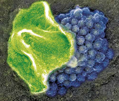
| As a pox virus fuses
with a cell it has just infected, it discards the shroud-like covering
it uses to “hide” as
it spreads from cell to cell within the body (below, green). The
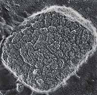
complicated tracks of protein-crystals seen on a “naked” pox
virus (right, without shroud) mix after fusion with the cell membrane,
making the collection of “mulberries” (below, blue).
The event is captured using “quick-freeze, deep-etch” electron
microscopy, a process created by John E. Heuser, MD, professor of
cell biology and physiology, which lets biologists take detailed
pictures of fleeting events inside living cells.
|
 |
 |