
|
|
||
|
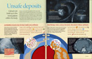
No Bones About It Calcium, the foundation of strong bones, deposits in arteries and elsewhere to devastating effects. But bone-making holds clues to thwarting vascular calcification.
|
||||||
|
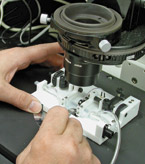
The pressure-diameter relationships of blood vessels can be directly measured using this precision apparatus. Thus, potential treatments that reduce or remove medial artery calcification can be evaluated and rated for how well they restore normal artery function. The calcified areas are — like bone — living tissues undergoing active remodeling. And that means the process of calcification could potentially be reversed. |
Calcium is a very good thing — in the right places. Bones contain most of the body's calcium, providing the body with a sturdy frame; the rest forms the teeth. But when calcium gets into blood vessels, the resultant hardening can threaten people's health. Dwight A. Towler, MD, PhD, director of the Division of Bone and Mineral Diseases, says that in some people, blood vessels and valves can become the second-most calcified structures in the body. Until recently, scientists and physicians knew very little about how calcium was deposited in blood vessels or how to get rid of it. Calcification of vascular components often develops with age and with disorders such as elevated cholesterol, high blood pressure, diabetes and kidney disease. While many of us are familiar with calcification of atherosclerotic plaques, those nasty lumps that can block arteries and contribute to clot formation, less well known is another common type of arterial calcification: medial artery calcification, or MAC. MAC describes calcium deposition within the matrix of artery walls and encircling the entire vessel. The hardening of the arteries associated with MAC interferes with the normal flexibility of blood vessels, which in turn impedes the proper flow of blood to tissues and puts extra stress on the heart. "Clinical data from populations susceptible to type 2 diabetes have shown that MAC is the single best predictor of risk for lower leg amputation and for mortality," says Towler, the Ira M. Lang Chair in Medicine and professor of molecular biology and pharmacology. "But I'm just old enough to remember being told that MAC isn't medically relevant because it doesn't block the artery like plaques can." Towler says MAC is largely unaddressed by available clinical treatments, even though the problem is widespread and serious. "I recently consulted on a patient with kidney failure whose blood pressure falls precipitously when she stands. In her X-rays, vessels appear with calcification all along them — you shouldn't be able to see blood vessels in X-rays at all. Because hers are calcified, they can't contract to compensate for the change in posture when she stands. The treatment options for such a patient are limited." Like many patients with arterial calcification, this woman also has osteoporosis. Physicians have noted that bone calcification and blood vessel calcification are often linked in a reciprocal relationship — as one goes down, the other comes up. This connection hints at something more complex in arterial calcification than previously realized. Earlier thinking held that calcification of arteries is a passive process; that is, calcium settles in blood vessel walls or deposits at places where cell death occurs. But Towler and colleagues have uncovered processes that actively create calcium deposits in arteries. One of the first discoveries on the way to this new understanding of vascular calcification was the finding that calcifying atherosclerotic plaques produced a powerful substance known to be involved in the repair of bone — bone morphogenetic protein-2 or BMP2. A growth factor, BMP2 was first identified at sites of bone formation and has since been successfully used to speed healing of broken bones. BMP2 promotes vascular calcification by activating Msx2-Wnt signaling, a bone-forming mechanism critical for skull and tooth calcification during embryonic development. A few years ago, Towler and colleagues performed experiments that marked another turning point in thinking about vascular calcification. They gave parathyroid hormone to laboratory mice to see how it would affect their arteries. Ordinarily, pulses of parathyroid hormone increase bone mass and bone strength; it currently is being used as a treatment for osteoporosis. To Towler's surprise, instead of increasing calcification in blood vessels of the mice, the hormone inhibited the vascular Msx2-Wnt signals that promoted arterial calcification — exactly the opposite of what occurred in bone.
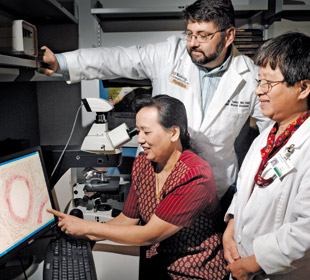
Jian-Su Shao, MD, research instructor in medicine, sitting, and Su-Li Cheng, PhD, right, research assistant professor of medicine, work with Dwight A. Towler, MD, PhD, studying the processes of vascular calcification. "Pulsatile parathyroid hormone signaling turns out to be an exquisitely smart hormonal rhythm," Towler says. "It directs the deposition of calcium in bones while simultaneously protecting blood vessels from calcification. We need to find out what mechanisms could account for that." The researchers found that blood vessels with MAC have gene activities commonly seen in bone and tooth formation. As Towler points out, if gene programs are turned on during calcification, that shows the process is definitely not passive. The reason patients can have osteoporosis along with artery calcification — why bones lose calcium as arteries gain it — lies in the fact that some cells in blood vessel walls can be molecularly "re-programmed" to become bone-making cells. These cells react in an opposite way from bone-making cells in the skeleton in response to the metabolic, hormonal and inflammatory changes that occur with diabetes, renal failure and advanced age. Investigations that followed these discoveries mapped out a detailed system of molecular signals involved in vascular calcification. Spurred on by high fat diets or diabetes, for example, cells in the outer layer of arteries begin producing BMP2. Reactive oxygen molecules and inflammatory substances produced by macrophages and fat cells play a role in initiating this step. Tiny blood vessels, or capillaries, within the walls of large arteries carry signal substances generated in reaction to BMP2. These signals combine to initiate a series of responses that transform arterial cells into bone-forming cells. The transformed cells use calcium to create an inflexible, brittle core within the vessel walls. A similar process leads to calcification of atherosclerotic plaques, but with the added contributions of cell death. "Physicians don't generally approach vascular medicine this way," Towler says. "They tend to think of what's happening in the layer of cells that lines the interior of vessels, where the blood flows. But we've found that it's the outer layer, the one furthest from the blood flow, where the important reactions are taking place that lead to calcification of the type seen in diabetes and kidney disease." This information about vascular calcification lends hope for effective treatments. It says that vessels aren't just absorbing calcium and turning to stone. Instead, the calcified areas of MAC are — like bone — living tissues undergoing active regeneration and repair. And that means the process could potentially be reversed — at least for this one type of vascular calcification. "If medial artery calcification is a disorder that responds to 'osteotropic' or bone formation hormones, such as parathyroid hormone, then it should be therapeutically tractable," Towler says. "If we take some of the things that we know about mineral deposition in the skull and long bone and begin to study those pathways in the vasculature, we may be able to inhibit it. That's an entirely new way of thinking about the problem."
|
|||||