
|
|
||
|
Origins of Alzheimer's Measuring A-Beta could lead to ways to abate it — and the onset of Alzheimer's.
|
||||||
|
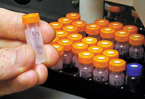
Aβ from human cerebrospinal fluid has been processed for measurement of the label by a sensitive device called a mass spectrometer, which can weigh the difference of a single neutron (smaller than an atom). The secret of the new test's success lies in a technique for temporarily tweaking brain chemistry that's so subtle it can only be detected by scientific equipment, not by brain cells or the chemical reactions that take place in the brain. |
Researchers have known since the early 20th century that a characteristic sign of Alzheimer's disease is that it leaves sufferers' brains riddled with plaques. The key ingredient of the plaques is a fragment of a protein known as amyloid precursor protein (APP). The fragment itself is called amyloid beta (Aβ). The causes of excessive Aβ are often maddeningly elusive. That's partially because a wealth of environmental and genetic factors probably contribute to risk. But another significant question also obscures scientists' view of Alzheimer's origins: Do patients' brains make more Aβ, or are they unable to clear it out quickly enough? A new test developed by researchers at the Alzheimer's Disease Research Center (ADRC) at Washington University in St. Louis may finally help resolve this mystery. The test is the first to allow researchers to safely monitor production and clearance rates of Aβ in humans. In addition to helping scientists learn more about the origins of Alzheimer's disease, the test may help them improve its diagnosis and treatment.
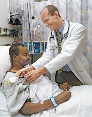
Through clinical studies with volunteers such as Carl Garrett, researcher Randall J. Bateman, MD, and colleagues are studying the body's production and clearance rates of the protein fragments that collect and impair the brains of Alzheimer's patients. It's very important to understand if patients have a decreased clearance rate or an increased production rate," says Randall J. Bateman, MD, assistant professor of neurology and lead developer of the new test. "If we find that it's mainly a problem in production or in clearance, then that's obviously what we want to be targeting with therapeutics." Practically speaking, one of the biggest obstacles to understanding Alzheimer's disease has always been the delicacy and inaccessibility of the living human brain. Neuroscientists generally aren't too eager to directly poke or prod it. Researchers had found ways to measure Aβ levels in the brain by taking samples of cerebrospinal fluid. But like many other proteins, APP, the precursor to Aβ, is regularly produced, used, discarded and reproduced, starting the cycle again. This made a reading of a patient's Aβ levels like taking a photograph of a sink flooding with water: It was useful to know that the sink was flooding, but they still couldn't tell if the faucets had been opened wider or the drain was clogging. The secret of the new test's success lies in a technique for temporarily tweaking brain chemistry that's so subtle it can only be detected by scientific equipment, not by brain cells or the chemical reactions that take place in the brain. In effect, it's a way of gently poking the brain without breaking anything. "The vast majority of the carbon atoms in our body are what we call carbon 12, meaning they have 12 protons and neutrons in their nucleus," Bateman explains. "We give patients an intravenous infusion of the amino acid leucine, a building block for proteins, that has been labeled with carbon 13, which has 13 protons and neutrons in its nucleus." The label is safe and non-toxic; rats have been raised entirely on food containing carbon 13, Bateman notes. All cells incorporate the labeled leucine into the proteins they make over the next several hours, including brain cells making APP. Scientists take periodic samples of the subjects' cerebrospinal fluid through a lumbar catheter, purify the Aβ from the samples and then use a mass spectrometer to determine how much of the Aβ includes carbon-13-labeled leucine. By tracking how fast the percentage of labeled Aβ goes up, they can determine the Aβ production rate. Bateman notes that the labeled leucine is very hard to separate out. "It behaves for all intents and purposes like the real thing, but that's why we chose it — because it doesn't interfere with the underlying physiology," he explains. After the labeled Aβ level peaks, researchers stop infusing the label but continue to regularly sample the cerebrospinal fluid. As old, labeled Aβ is cleared away and cells no longer have the labeled leucine to incorporate into new Aβ, the percentage of labeled Aβ goes down, revealing its clearance rate. Bateman developed the mass spectometry technique with extensive guidance and assistance from Kevin E. Yarasheski, PhD, associate professor of medicine and assistant director of the Washington University Biomedical Mass Spectrometry Resource, and staff scientist Ling Munsell, M.S. Yarasheski previously used a similar approach to measure production and clearance rates of muscle proteins. Working in the laboratory of David M. Holtzman, MD, the Andrew B. and Gretchen P. Jones Professor and head of the Department of Neurology and with the support and guidance of John C. Morris, MD, the Harvey A. and Dorismae Hacker Friedman Distinguished Professor of Neurology and director of the ADRC, Bateman tried out the new test on six healthy, young volunteers. Because Alzheimer's symptoms take many years to develop, some researchers had assumed that the creation and clearance rates for Aβ were very slow. But the initial test of the new technique suggested the opposite. "Amyloid beta has the second-fastest production and clearance rate of any protein whose production rate has been measured so far," Bateman says. "In a time span of about six or seven hours, you make half the amyloid beta found in your central nervous system." As expected, in the first six healthy volunteers, the clearance rate closely matched the production rate. "Without a balance in those two rates, a person will end up with increasing or decreasing amounts of amyloid beta in the brain," Bateman notes. Bateman and his colleagues have begun applying the test to Alzheimer's patients to determine whether their production or clearance of Aβ is changing. There's also substantial potential to apply the new test in clinical contexts. Previously, the only way to assess the effectiveness of a new Alzheimer's drug was to follow the mental performance of patients receiving the treatment over many months to years. "This new test could let us directly monitor patients in clinical trials to see if the drug is really doing what we want it to do in terms of Aβ metabolism," Bateman says. "If further study confirms the validity of our test, it could be very valuable for determining which drugs go forward in clinical trials and at what doses." The test also may be useful in diagnosis of Alzheimer's prior to the onset of clinical symptoms, which occurs only after Alzheimer's has inflicted widespread and largely irreversible damage on the brain. "One of the problems with Alzheimer's disease \and most dementias is that once they set in, even if you stop the offending process, the brain doesn't necessarily reverse and heal itself to its normal state," Bateman explains. "We may be able to use a modified version of this new test to detect a metabolic imbalance in amyloid beta before people become demented, and start treatment then." The technique may have applications beyond Alzheimer's disease. Other research groups have expressed interest in applying the new test to neurological disorders such as Huntington's disease, Parkinson's disease and prion protein diseases.
|
|||||