
|
|
||
|
Measured Impulses Understanding the electro-physiological properties of the heart can help prevent a leading cause of sudden death
|
||||||
|
This array of sensors offers dramatic improvement over the traditional electrocardiogram, according to Yoram Rudy, PhD, director of the Cardiac Bioelectricity and Arrhythmia Center. The technology offers the means to pinpoint electrical trouble spots in the heart. Related Links
Rudy's Lab
CBAC
|
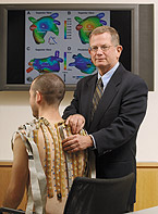
… KA-thump. KA-thump. KA-thump. KA-thump … "Sometimes you hear about an athlete who falls dead on the basketball court or an older man who survives a heart attack and then later dies unexpectedly while driving his car," says Yoram Rudy, PhD, the Fred Saigh Distinguished Professor of Engineering. "These sudden deaths are likely due to rhythm disorders of the heart. Arrhythmias cause disability or a com-promised quality of life for many more people, and the incidence of these disorders is increasing as the population ages." Rudy came to Washington University in 2004 and set up the Cardiac Bioelectricity and Arrhythmia Center (CBAC), which he directs. CBAC brings together researchers from the Danforth and medical campuses, including biophysicists, physiologists, biomedical engineers, cardiologists, radiologists and surgeons. CBAC members are uncovering new information about the electrical properties of the heart and the causes of cardiac arrhythmias using mathematical and computational tools and novel imaging technologies. Arrhythmias occur when heart defects perturb the electrical impulses responsible for the heart's contractions. Electrocardiogram, or ECG, a more than 100-year-old technology, measures these electrical signals using electrodes attached to a patient's skin. ECG can diagnose arrhythmias and monitor the effects of drugs or devices for regulating the heart, but relies on just six to 12 electrodes. Rudy foresaw that a more precise method for measuring the heart's electrical activity would be a significant medical advance. He and his colleagues ultimately came up with a vest-like array of 250 electrodes that allows highly detailed mapping of electrical signals emanating from the heart. Just as importantly, they found ways to combine these readings with computed tomography (CT) images of the heart to pinpoint the source of abnormal electrical activity. This new technology is called electrocardiographic imaging or ECGI.
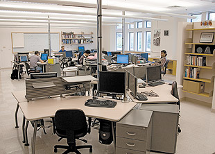
The CBAC computer lab's open floor plan facilitates collaboration. Predicting the heart's electrical activity based on signals picked up from the skin requires working backwards from effect to cause; researchers term this "the inverse problem." "Not only did we need to devise the mathematics to solve the inverse problem, we had to deal with the fact that the signal at every body-surface electrode is an integral effect of the electrical activity over the entire heart," says Rudy, also professor of biomedical engineering, cell biology and physiology, medicine, radiology and pediatrics. "Then we had to develop computer algorithms to combine the electrical measurements with realistic geometries of the human torso and heart." School of Medicine researchers are collaborating in CBAC to test ECGI as a diagnostic and treatment tool. A proof-of-concept study of ECGI for treatment of children with Wolff-Parkinson-White syndrome (WPW) showed how quickly the technology could locate problem areas on the heart. "Kids with WPW have a short circuit in their heart muscle, says Edward K. Rhee, MD, adjunct assistant professor of pediatrics at the School of Medicine and director of Invasive and Arrhythmia Services at St. Joseph's Hospital and Medical Center in Phoenix AZ. "The standard mapping procedure takes about two hours; with ECGI, it was done in minutes." In the standard procedure, the treating physician has to move a mapping catheter bit by bit through the heart to map the electrical activity of the whole organ. ECGI was able to find the source of the problem in the hearts of children with WPW just as accurately, but in a much shorter time and without the need for an invasive procedure. Rhee asserts that one promising future application of ECGI is treatment of a type of heart failure in which areas of the heart contract too late. ECGI can guide placement of pacemaker electrodes, which then resynchronize contractions. Bruce D. Lindsay, MD, professor of medicine and director of the Clinical Electrophysiology Laboratory, is a member of CBAC. Along with other types of arrhythmias, Lindsay investigates and treats atrial fibrillation in which the heart's upper chambers contract chaotically. "ECGI can help us decide what areas of the heart to target with ablation techniques to eliminate fibrillation," Lindsay says. "But it also promises a better fundamental understanding of the circuits that cause atrial fibrillation and the regions of the heart that sustain the abnormal contractions." If the technology can be developed so that patients can wear the ECGI vest while going through their typical day, it could record electrical activity during arrhythmic episodes and would be far more sophisticated than anything now available, according to Lindsay. ECGI's high resolution has potential to help surgeons better plan antiarrhythmic heart surgery, and some day its precision also may help doctors screen and identify patients at high risk of sudden death before it's too late. But ECGI isn't the only implement in the CBAC toolbox. The researchers also developed computer models that replicate the complex functions of heart muscle cells to learn more about the mechanisms underlying cardiac arrhythmias. The heart's electrical impulses are carried by charged molecules, or ions, as they flow through channels in the walls of the heart's cells. Minute changes in the structure or other properties of these channels — through genetic mutation or disease — can interfere with ion movement and disrupt the heart's electrical cycle. Rudy's group developed mathematical formulations to account for the various ions and channels in real cardiac cells. Using their computer models, researchers can manipulate virtual heart cells to test the effect of alterations in ion flow and channels. Because the heart's cells work together, Rudy's lab has taken computer modeling to the next step and combined cells to recreate virtual cardiac tissue. Their cellular and multicellular computer models can be used to identify drug targets, steer drug design and simulate the effects of arrhythmia treatments. Together, ECGI and cardiac computer modeling represent a successful wedding of basic research and theory to clinical practice. "Within CBAC, we are working closely with a variety of researchers and clinicians — in pediatric cardiology, adult cardiology, cardiothoracic surgery and radiology," Rudy says. "We are so pleased to be taking our basic research to research and application in people. We are now starting to have an impact on the medical treatment of arrhythmias."
|
|||||