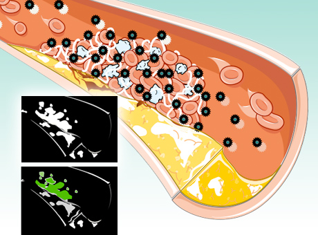


For almost two decades, cardiologists have searched for ways to see dangerous blood clots before they cause heart attacks.
Now, researchers at the School of Medicine report they have designed nano-particles that find clots and make them visible to a new kind of X-ray technology.
According to Gregory M. Lanza, MD, PhD, a Washington University cardiologist at Barnes-Jewish Hospital, these nanoparticles will take the guesswork out of deciding whether a person with chest pain is actually having a heart attack.
“Every year, millions of people come to the emergency room with chest pain. For some, we know it’s not their heart. But for most, we’re not sure,” says Lanza, professor of medicine. When there is doubt, the patient must be admitted to the hospital and undergo tests to rule out or confirm a heart attack, costing money and time.
This new technology could reveal the location of a blood clot in a matter of hours.
The nanoparticles are designed for usewith a new type of CT scanner that “sees” metals in color. The new technology, called spectral CT, uses the full spectrum of the X-ray beam to differentiate objects that would be indistinguishable with a regular CT scanner that sees only black and white.
In this case, the metal in question is bismuth. Dipanjan Pan, PhD, research assistant professor of medicine, designed a nano-particle that contains enough bismuth for it to be seen by the spectral CT scanner.
More than simply confirming a heart attack, the new nanoparticles and spectral CT scanner can show a clot’s exact location.
With this imaging technique, Lanza predicts new approaches to treating coronary disease, technologies that might act like Band-Aids, sealing weak spots in vessel walls.
“Today, you wouldn’t know where to stick the Band-Aid,” Lanza says. “But this technique would show the exact location of clots in the vessels, making it possible to prevent the dangerous rupture of unstable plaque.”