
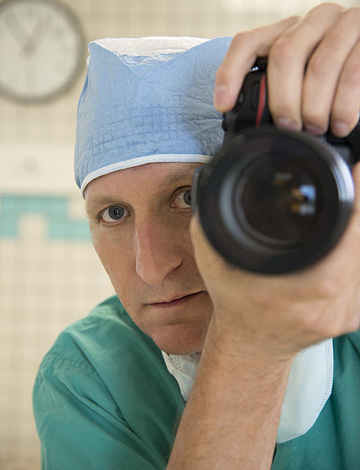
ROBERT BOSTON


It is a sacred place, a place to acquire vast amounts of knowledge about the human body. A space few people get to see. These are the reasons I chose the Anatomy Dissection Lab as the subject for my photo essay.
First-year medical students are introduced to the human body immediately in the dissection lab. It is the entry way to the study of medicine, a rite of passage that all medical students experience on their way to becoming a doctor.
I started photographing the dissection lab late last summer before the 120 students began their studies. This allowed me to move around more easily while photographing the glass-encased human body specimens lining the walls. Many are more than 100 years old. Virtually every part that makes up the human body is on display for students to view. Many were beautiful to me, while others were strange and disturbing.
I photographed alone for the first few days. I was fascinated and drawn in. The purple heart in a square jar was mesmerizing. The greyish-green organ in a large rectangular jar looked very fragile. I thought about the organs working in my own body. Are my parts working OK? Is everything synchronizing correctly in me? The whole thing seemed impossible. Amazing, intricate, ornate, grotesque, gorgeous and scary are some of the words that passed through my mind as I photographed. Where do you order those jars from anyway?
It was difficult to choose which objects — in jars and under glass domes — and freestanding skeletal parts to focus on. There were too many visually overloading photographic possibilities. To overcome this dilemma, I subtracted. Artists, and especially photographers, subtract to simplify a scene. Take only certain things you want to see in the frame and leave out the rest. This day I chose to shoot the body parts that had the most interesting shapes. Shiny chrome dissection tables, which cadavers would eventually lie on, also caught my eyes.
As I worked, the air seemed to become heavier and heavier. After three hours, I became exhausted, quit for the day and headed back to the office. Photographing in the operating room, emergency room and Intensive Care Unit can be tiring, but this feeling of tiredness was different. Was it because the body parts are basically the same parts that are in me, but strange to see on display? I wondered if it was just me, or whether the lab is a difficult place for everyone at first and eventually becomes easier … not unlike getting used to cold or hot weather. The cadavers are not in the room yet!
On the third day of shooting, the embalmer instructed me to come downstairs and retrieve the keys that open the glass cases. I took the elevator down to the morgue. It was the most organized space I had ever seen. Everything was in its place on shelves and spotless. This is where each donated body is prepared for the students to eventually study.
On day four, I finished photographing in the quiet lab. Soon the students, instructors and cadavers would occupy the space. I originally planned to include only the anatomical specimens on display and the lab space. My photo essay would be incomplete had I done this. It needed to include the room with instructors and students as dissection took place.
The fifth and last day of shooting was on the second week of the anatomy class. The room was filled with students dissecting and studying human bodies under the direction of four faculty members. It was quite a different experience. I entered the room and tried to be inconspicuous. How was that going to work? I was the only one with a camera and no scrubs. It was one of the few shoots where I never became invisible.
Dr. Glenn Conroy, the course master, asked me to not include much of the cadavers in my photographs, out of respect to the donor project. This was my intention anyway, but I didn’t realize how difficult it would be.
It was a challenge to capture the scene artistically and informatively without the inclusion of cadavers. Most of the students were calm and focused. Others seemed disturbed and nervous with the experience and understandably so. All had never experienced anything like this. Dissecting a frog in high school was not a sufficient prerequisite. Instructors — familiar with dissecting the human body — guided students through the process, or journey, if you will.
Writing allowed me to look back at this project. Most of it was enjoyable, some of it not so much. All of it was a learning experience. I found the anatomy lab to be a place of energy, intensity, knowledge and respect.
Special thanks to Dr. Glenn Conroy for allowing me to photograph in the dissection lab.
