

The Shape of Things to Come
Through a delicate procedure that is part surgery and part sculpture, Judith M. Gurley, MD, helps a baby with a cranial deformity get headed in the right direction
The reconstructed skull is a patchwork quilt of pieces, with gaps in between. Within three months, the skull bone will grow back and fill in these spaces.
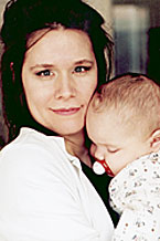
Judith M. Gurley, MD, with her young patient, Brian Bauman.
![]()
Soon after Brian Bauman was born, his parents heard the chilling news that something was amiss. The shape of his head was wrong—perhaps a result of his difficult birth—but might correct itself with time. Instead, Jeff and Karen Bauman of Granite City, Illinois, watched day by day as their son's abnormally narrow skull bulged more visibly in the forehead and in the back.
Their pediatrician sent them to St. Louis Children's Hospital, where doctors diagnosed sagittal synostosis, a condition that occurs once in every 4,000 to 5,000 births. Brian's cranial bones had fused prematurely, so he lacked the usual infant “soft spot.” His brain, restricted from growing side to side, was forced to push forward and backward, giving his head an elongated shape. Left untreated, Brian eventually might suffer from headaches, and his skull deformity would worsen. He would have a hard time lying on the back of his head and wearing a hat or bike helmet.
Surgery was the only solution, but the prospect made the Baumans anxious. Their older son, Brandon, now a healthy 4-year-old, was born prematurely and had numerous medical problems. A second son, Blake, had died when he was six weeks old after an Illinois clinic misdiagnosed his bronchial pneumonia as an allergic reaction.
Still, the family decided to go ahead with the delicate procedure in which a neurosurgeon would peel back Brian's scalp and remove his skull piece by piece. Then a plastic surgeon would reshape those pieces before reattaching them to the remaining skull. “It is just something that has to be done. We don't want him to suffer,” says Jeff Bauman, a union carpenter.

The shape of baby Brian's head, before (white) and after the surgery that reshaped his skull.
For the surgery, the family turned to Judith M. Gurley, MD, assistant professor of plastic and reconstructive surgery, who joined the Washington University faculty in 1999—one of a talented cadre of women who are breaking new ground in traditionally male surgical subspecialties. In fact, when Gurley first came to the School of Medicine in 1998 for a pediatric craniofacial fellowship, she was one of a few American women in her field.
At her first meeting with the Baumans, Gurley explained the risks and benefits of surgery. Although she and her partner, Jeffrey L. Marsh, MD, professor of plastic and reconstructive surgery, have performed dozens of similar procedures—10 per year on average—without incident, there is a small risk of serious complications. A few centers have reported fatal bleeding incidents; every child who undergoes the procedure needs a blood transfusion.
To minimize risks and achieve the best cosmetic result, children with sagittal synostosis should have the surgery as early as possible. Infant skull bones are malleable, so the surgeon can use special scissors and bone-bending forceps to re-form the pieces.
“The older the child is the more difficult the surgery, because you can't bend the bone and you need a saw to shape every piece,” says Gurley. “That increases the operative time, increases the blood loss and makes it a much more laborious procedure.”
Surgery Day Arrives
So on October 30, a few days shy of his four-month birthday, Brian Bauman and his parents are in a waiting room at Children's Hospital. At 17 pounds, he is a husky baby— and since midnight he has not been allowed to eat. Now it is nearly lunchtime, and he is beginning to whimper. His mother soothes him. Brandon is not with them; he is back home feeling “scared and angry,” his mother says, because he remembers Blake's death—and fears the same thing will happen to Brian.
With cheerful words of reassurance, anesthesiologist Julian Yepes, MD, whisks Brian away to the operating room. Yepes will spend one to two hours anesthetizing the baby, hooking up his intravenous lines, placing a catheter in his bladder, inserting a breathing tube and swaddling him in a heating blanket that will keep him warm during surgery. Then the stage is set for neurosurgeon Jeffrey G. Ojemann, MD, assistant professor of neurological surgery, to begin his part of the procedure.
In the blue-and-white world of the operating room, any other color stands out sharply. On this day before Halloween, several members of the OR team—which also includes a scrub nurse, circulating nurse and neurosurgery fellow Tord D. Alden, MD—wear jaunty orange headgear.
To begin, Ojemann makes an ear-to-ear incision. Working forward and backward, he peels away Brian's scalp, exposing his skull from his eyebrow bone to the back of his neck.
On the exposed skull, he draws a pattern of lines and dots that will provide a blueprint for his cuts. He starts his saw, which is tethered to a foot pedal; whining like a dentist's drill, it slices neatly through the skull bone. Brian's forehead bone comes off first, then others—four in all. What remains is a strip down the middle of Brian's skull and a section above each ear, where the reshaped pieces will later be reattached.
The process, which looks routine in Ojemann's hands, is actually fraught with risks. The surgeon has to saw through bone without disturbing major blood vessels or the brain's protective lining—a process made easier by the saw's safety features. Still, Ojemann says, “in such a small child, even a small blood loss can be fatal, so we must make absolutely sure the lining is free from the bone. We take great pains to do that before we make our cuts.”
Reshaping the Skull
When Ojemann is done, Gurley and her surgical assistant, craniofacial fellow Delora Mount, MD, are summoned to the operating room for the next phase of surgery. The procedure they will use is only three decades old, developed by French plastic surgeon Paul Tessier, MD. Studying each section of Brian's skull in turn, Gurley squeezes, snips, bends and stretches, casting occasional glances at the CT-scan posted on the wall.
This bone manipulation gives Gurley an affinity with orthopaedic surgeons. “In fact, a lot of people call craniofacial surgery 'orthopaedic surgery of the face,'” she says. “I have always liked working with bone, since human form is largely dependent on the bone that supports our soft tissue. And I enjoy making changes that are visual.”
That aesthetic sense gives Gurley another kind of kinship: with painters, sculptors and other artists. As a University of North Carolina undergraduate, she took figure drawing classes; today, she still paints in her spare time. Medicine and art intersect in plastic surgery, which brings the two together in “the perfect combination,” she says.
But the experience that clinched Gurley's interest in pediatric craniofacial surgery occurred during her residency at the University of Chicago, when she twice traveled to Central America with surgical teams on missions of mercy. There, she had the chance to operate on desperately poor children long overdue for medical care.
Today, she is hard at work on tiny Brian Bauman: inspecting each reshaped piece of skull, then holding it up to his head with an appraising look to see how it will fit. Using a surgical saw, she also makes barrel-stave cuts in his eyebrow bone and in the portion of skull above each ear. These cuts will allow the constricted brain to reshape itself, expanding side to side. Finally, she begins attaching the skull pieces to these serrations with dissolving plates and screws.
The reconstructed skull is a patchwork quilt of pieces, with gaps in between. Within three months, the skull bone will grow back and fill in these spaces. Meanwhile, this extra room will give the brain more breathing space, but it will also make Brian's head more vulnerable. A week after surgery, Brian will begin wearing a helmet for protection until his bone has fully regrown.
Just after 5 p.m.—little more than five hours after it began—Brian's surgery is nearly finished. Gurley pulls his scalp back up and deftly stitches it together. She wraps his head in gauze, as he has lost a good deal of blood and more will drain. The scar will be watertight within 48 hours. The baby will only need to spend three days in the hospital.
“Isn't he beautiful?” asks Gurley, admiring Brian's skull. Next she will hurry out to talk to his waiting parents. The mother of a toddler herself, she can imagine how they feel.
“It is fascinating to see parents before and after surgery,” she says. “Beforehand, no matter how gently you describe the procedure, they are petrified. Afterwards, there is such a sense of relief—and every single one has been glad they did it.”
Follow-up Visit
Early in January, Brian is back at Children's Hospital for his two-month visit. His head measurements are perfect, Gurley says, and with his chubby face and round skull, he has a whole new look. “It's amazing,” says Karen Bauman. “He looks like his dad now — he never did before.” Their older son Brandon is relieved but still protective, she adds. “He'll tell me, 'Mom, watch his head.'”
The only vestige of surgery is a thin pink scar running across the top of Brian's head, from one small ear to the other. Already, his downy blond hair is covering it up, and soon it will be hidden completely.
If he ever goes bald, that old scar may one day reappear.
But otherwise, Brian Bauman will never have any reason to recall the extraordinary
surgery that changed his appearance and his life. ![]()