
|
|
||
|
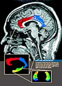
What's Inside Schizophrenia? One in one hundred young minds may develop schizophrenia.
|
|||||||
If people who don’t have schizophrenia have similar brain anatomy to their siblings who do, they may be at risk.
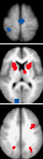
Control subjects show cognitively related brain activation in the red areas; individuals with schizophrenia do not. Conversely, schizophrenic individuals show greater brain activity in the blue areas, compared to controls. |
IMAGINE YOU HAVE JUST BEEN DIAGNOSED WITH SCHIZOPHRENIA. If you’re a typical patient, you are a 19- or 20-year-old man or a 21- to 22-year-old woman, and you’ve probably had active psychotic symptoms for about a year and a half. Maybe it was an individual voice or a peculiar thought at first. But that single, isolated symptom grew. The voices became louder and more numerous, the delusions more elaborate. Soon, you found yourself in the emergency room or clinic, and the diagnosis of schizophrenia was made. For most patients, that’s when treatment begins. John G. Csernansky, MD, would like it to start sooner. He’d like to find a way to prevent that first episode. "As valuable as it is to understand how to prevent subsequent episodes, the key to really changing outcomes is to begin treatment before the first episode occurs," he says. "We need to find a way to predict who is going to become psychotic and treat them before they do." Just less than one percent of the general population suffers from schizophrenia. After that first event and diagnosis, chances are that patients will relapse—especially if they stop taking medicine. "Generally a second psychotic episode lasts longer and is more severe," Csernansky says. "It also can be more difficult to treat." And each relapse tends to get a bit worse. Somewhere between 10 and 15 percent of patients have frequent
relapses. Often, they end their lives in institutions suffering with chronic
psychosis. Even the best possible outcome—no future relapses or psychosis
—still requires patients to take antipsychotic medications for the
rest of their lives. After the initial event, something has happened in
their brains that cannot be undone. Csernansky, the Gregory B. Couch Professor of Psychiatry and associate professor of anatomy and neurobiology, spent the last half of the "Decade of the Brain" looking for ways to identify anatomical differences in the brains of patients with schizophrenia. Now, thanks to a grant named for the man who sponsored the congressional resolution that designated the 1990s as the "Decade of the Brain," he’ll get a chance to expand that work. Last fall, the National Institute of Mental Health awarded a three-year, $2 million grant to launch a Silvio Conte Center at Washington University. Csernansky, the new center’s director, will oversee several major projects that are attempting to locate and identify differences in the brains of patients with schizophrenia and similar psychiatric disorders. MAPPING THE BRAIN This pursuit began in 1995 as a collaboration between Csernansky’s group in the School of Medicine’s Department of Psychiatry and Michael I. Miller, PhD, who was then in the Department of Electrical Engineering on the university’s Hilltop campus. Miller now is a professor of bio-medical and electrical engineering in the Whiting School of Engineering at The Johns Hopkins University in Baltimore. There he develops algorithmic metrics in the emerging field he terms "computational anatomy" that allow him to capture and measure the shapes and sizes of biological and anatomical structures. In the context of the Conte Center brain mapping project, Miller develops computer algorithms that take brain scans and convert those two-dimensional images into three-dimensional models. Called high dimensional brain mapping (HDBM), the technique allows scientists to spot differences that might contribute to problems. "You might compare the technique to tracing a pile of pictures of faces," Miller says. "If the pictures were stacked one on top of another so that all of the facial features lined up, we could trace a ‘standard’ face and then compare individual variations to that standard template. Our algorithms do the same kind of thing with the brain."
John G. Csernansky, MD, right, discusses results with Lei Wang, PhD, research associate in psychiatry, and Deanna M. Barch, PhD. Imaging systems and sophisticated mapping software help reveal the characteristics of the schizophrenic brain. When the HDBM computer programs have worked their magic, it’s possible for researchers to spot tiny differences in the size and shape of brain structures. Csernansky compares it to looking at a damaged car. "In the past, the limitations in our imaging technology made it necessary for the car to be missing a door or an entire fender before we could spot a difference," he explains. "What Mike Miller’s group has given us is the ability to see scratches in the paint or hail damage, things that were undetectable before." The idea that there may be anatomical differences in the brains of people with schizophrenia is not a new one. Since the 1970s, some neuroscientists have argued that such changes might contribute to schizophrenia and other mental illnesses, but the complexity of brain mapping and the amount of variation between individuals made it nearly impossible to prove. Until now. Using Miller’s computer algorithms, Csernansky has identified small but distinct deformities in shape in the hippocampus and other brain structures, even in newly diagnosed patients. As research progresses at the new Silvio Conte Center, the investigators hope to identify other differences between the brains of patients with schizophrenia and those who don’t have the disease. IDENTIFYING RISK Working with C. Robert Cloninger, MD, the Wallace Renard Professor of Psychiatry and professor of genetics at the School of Medicine, Csernansky will conduct imaging studies of patients just diagnosed with schizophrenia, their younger siblings, and age-matched controls. Taking advantage of Cloninger’s expertise in genetic influences on psychiatric illness, the project will use detailed family histories to determine whether siblings of some patients might have a genetic risk of developing schizophrenia. In theory, those at risk might have the same anatomical differences as their affected siblings. By making brain scans, the researchers hope to find anatomical markers related to risk. If people who don’t have schizophrenia have similar brain anatomy to their siblings who do, they may be at risk. The presence of some abnormal anatomy in siblings may help determine whether some structural changes are a coincidence or whether they actually contribute to the development of schizophrenia. Cloninger and Csernansky will focus on a particular group of brain structures—those of the temporal lobe and the frontal lobe and the pieces that connect them. In particular, they will look for abnormalities in the thalamus, a part of the brain important to the exchange of information between the two lobes. While Csernansky’s team seeks anatomical abnormalities, a second project will look for functional differences. Using functional magnetic resonance imaging (fMRI) scans, a team led by Deanna M. Barch, PhD, assistant professor of psychology, and Randy L. Buckner, PhD, associate professor of psychology in Arts and Sciences, will ask subjects to perform a variety of tasks while in a scanner to learn whether patients with schizophrenia use their brains in different ways than healthy subjects. The fMRI scans reveal changes in brain activity. Barch and Buckner hope to learn how different types of memory are affected by schizophrenia and how the brains of patients with the disease function differently—especially in areas such as the prefrontal cortex, the hippocampus, the anterior cingulate and the thalamus. "We expect these tasks will demonstrate that schizophrenia is associated with specific changes in how well people perform both working and episodic, or long-term, memory tasks," Barch says. "In both areas, we expect individuals with schizophrenia will not do as well as healthy controls. We also would predict reduced functional activation—or at least altered activity—in the brain structures that we’ll be looking at. We hope to learn whether the unaffected siblings also have reduced behavioral performance and altered activity in these brain regions." If some siblings don’t do as well on the tests, or if their brains function in similar ways to their siblings who have schizophrenia, it could mean that they also are at risk for developing schizophrenia. TESTING AN ANIMAL MODEL A third Conte Center project will attempt to verify the results of the structural and functional imaging studies using animal models. Researchers at Yale University have developed a model of schizophrenia by exposing pregnant animals to X rays during the time at which brain cells in the thalamus are formed. Because they know the animals have damage in the thalamus,
the researchers will be able to compare the animal scans to human scans
of the thalamus to determine if similar anatomical abnormalities exist. "They exhibit an adult-onset deficit in the ability to perform a spatial memory task that is generally thought to reflect function in the prefrontal cortex," says Lynn D. Selemon, PhD, associate research scientist in neurobiology at Yale School of Medicine. "This is interesting to us because the onset of behavioral changes and cognitive decline in schizophrenia generally occurs in late adolescence or early adulthood. When they were younger, these animals performed this task as well as normal infants, but as adults, those who were exposed to radiation don’t do as well." As time goes on, Csernansky hopes to add more projects,
but at present, he hopes the new Conte Center can determine whether structural
and functional brain changes can predict risk of schizophrenia and, if
so, whether it will be possible to intervene.
|
||||||