
|
|
||
|
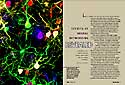
Secrets of Neural Networking Revealed Individual neurons stand out from the nervous system crowd
|
|||||||
"We are pushed to try new methods in the hope of getting
a better glimpse of this enigmatic system."
"In biology, like football, there’s a huge amount
of uncertainty in interpreting an image if you don’t know what happened
immediately before and after your snapshot." |
LIKE A COMMUTER NAVIGATING RUSH HOUR TRAFFIC, each nerve cell, called a neuron, must find its correct destination in the chaos of the developing brain. But unlike the commuter, each neuron also projects branches to multiple sites. The neuron faces the added challenge of deciding which connections to maintain and which to eliminate. "What’s fascinating about the nervous system is that not only are there such a vast number of connections, but somehow they all figure out the correct patterns of connectivity," says Joshua R. Sanes, PhD, the Alumni Endowed Professor of Neurobiology. He is one in a consortium of four highly collegial anatomy and neurobiology researchers who have been trying to uncover how cells know where to latch onto other cells and when to detach—a challenge recently made easier by the burgeoning field of optical imaging. With billions of neurons, each projecting a multitude of spindly "arms," it is difficult to pick out a single neuron from the crowd, particularly within the complicated context of an intact brain. But new techniques in optical imaging and molecular biology allow researchers to color and illuminate individual cells and, for the first time, watch interactions between neurons in their natural context, a living animal. "The big revolution in cellular and molecular biology is the ability to label living cells," says professor Jeff W. Lichtman, MD, PhD. "This is the birth of a new field which, in my view, will improve the quality of virtually all biology-related pursuits from physiology to chemistry." The School of Medicine team has developed and successfully launched these new tools in the laboratory. Now they are taking them one step further: In addition to watching neurons, researchers are using optical imaging and its results to manipulate cellular activity and isolate the genes responsible for each stage of the connectivity process. And, if none of the existing techniques can answer a particular research question, they invent a new one.
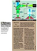
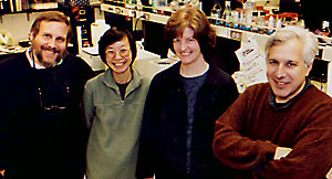
A collaborative team in developing new means to probe the mysteries of neural connectivity: Joshua R. Sanes, PhD; Rachel O.L. Wong, PhD; Ann Marie Craig, PhD, and Jeff W. Lichtman, MD, PhD. In the same way that ultrasonics uses sound, optical imaging employs light as a means of capturing an image. In the late 1990s, biologists discovered that the protein that makes certain jellyfish luminescent also could be used to generate fluorescent cells in other species. Soon, four variations had been developed, each one emitting a different color—green, red, yellow and cyan (greenish-blue)—when excited by light. By shining a light on a living transgenic mouse engineered to contain the genes that encode these jellyfish fluorescent proteins (FPs), researchers can watch cellular activity over time through a microscope. Peering inside living animals requires new kinds of imaging devices. Fortunately, two promising tools—confocal and two-photon laser microscopes—had just been developed when researchers began to experiment with FPs in mammals. These sophisticated optical microscopes enable researchers to capture three-dimensional images of biological structures deep within an animal, without damaging the tissues of interest. Such techniques are particularly valuable to neuroscientists, who have the daunting task of understanding what is arguably the most intricate, complex and sensitive organ in the body—the brain. "Neurobiology has been the frontier for the development of many new techniques," says Lichtman. "Because we still know so little about the workings of the nervous system and it is so difficult to study, we are pushed to try new methods in the hope of getting a better glimpse of this enigmatic system." THE CELLULAR / MOLECULAR CONNECTION Neurons communicate with each other across small gaps called synapses. According to Lichtman, a standard approach used to deduce how synapses form and mature—comparing images of different synapses at various developmental stages—is like a foreigner trying to figure out the rules of American football by putting together a sequence of still photos from various games. "In biology, like football, there’s a huge amount of uncertainty in interpreting an image if you don’t know what happened immediately before and after your snapshot," he says. As one of a few groups in the country studying neurons in living animals, the Washington University researchers were primed to test fluorescent proteins. Lichtman, Sanes and postdoctoral student Guoping Feng, PhD, were the first to show that FPs successfully label whole neurons in living mice without harming the fluorescent cells or the animals. To their surprise, in some of the transgenic lines of mice, the FPs labeled highly specific subsets of neurons. By mating mice with different labeling patterns, the team produced animals with only a few neurons labeled one color on a background of nerve cells labeled another color, providing a clearer picture of the connections between individual cells. The team uses these fluorescent mice to study the most accessible mammalian synapse, the neuro- muscular junction (where neurons connect to muscles). They already have discovered that neurons touch fewer and fewer muscle cells during the first few weeks of life, proving the theory that synapses are rapidly eliminated during development. Optical imaging allows them to watch in high resolution as neurons compete with each other to determine which branches disconnect from synaptic targets and which persevere. The use of FPs to study cell development is an example of how cellular biology has benefited from molecular biology: By using the latter’s techniques to identify a specific gene responsible for fluorescence in jellyfish and adapting that gene for use in mammals, scientists now can examine changes in the structure of mammalian neurons and other cells. In turn, FP-assisted cellular observations are contributing to the advancement of molecular pursuits. Sanes has identified several genes important for the development of initial synapses using more traditional molecular methods. But those techniques can only measure the end point—whether or not a healthy synapse formed. They cannot isolate which step of the process requires a given gene. Using Sanes’ transgenic mice, Sanes’ and Lichtman’s laboratories now can take a closer look at the neuromuscular junction. They mate the fluorescent-stained mice with those that lack a suspect gene. The resultant offspring have a few fluorescent neurons that, at some point during development, will not proceed as expected. The group monitors the neurons to detect when the glitch occurs, thereby obtaining a better understanding of the gene’s role.
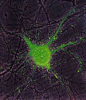
The bright green spots along a hippocampal neuron's branches reveal its many synaptic links, as captured and isolated by F. Thomas Crump, PhD, and Ann Marie Craig, PhD. INTO THE BRAIN The new optical imaging tools not only work well in the study of the neuromuscular junction, they also provide a unique opportunity for examining synapses in the brain. Associate professor Ann Marie Craig, PhD, investigates synapse formation between nerve cells taken from the hippocampus, a small structure in the center of the brain. For example, Craig grows hippocampal nerve cells in a dish and watches how they make, break and modify synapses. By attaching FPs to receptors that sense chemical signals exchanged between neurons, her group, working with Wong and Lichtman, follows the movement of FP-labeled receptors across synapses. Rachel O.L. Wong, PhD, associate professor, also studies the brain’s synapses, though in a slightly more accessible region—the eye’s retina. To study how developing neurons communicate with each other, Wong needs to simultaneously label many cells in the living retina and monitor their activity. Teaming with Wen-Biao Gan, PhD, and Jaime Grutzendler, PhD, postdoctoral fellows in Lichtman’s laboratory, Wong developed a new approach to rapidly label living cells with many colors: By using a "gene-gun" in a new way, tiny metal particles coated with different color dyes are delivered to the tissue, instantly labeling neighboring cells and their branches with one of seven colors. This technique, called DiOlistics, paved the way to study potential interactions between nearby neurons as they contact each other. Together with Gan, Wong also helped to pioneer a related technique called Calistics. Using dyes that sense calcium levels inside the cell, Wong can watch signaling between neurons in the retina as they form and establish contacts during development. Because connections between the many types of neurons are highly specific, Sanes and Wong believe that retinal neurons have different genetic makeups that affect their choice of synaptic partners. They are testing this idea by using Sanes’ FP mice to determine which genes are active in certain cell types. These genetic profiles are obtained using relatively new cDNA micro-array technology. According to Wong, the team’s fusion of new scientific tools and varied research perspectives is a creative means toward the more important end—understanding how the nervous system makes accurate connections during development. By employing these new techniques and collaborating to pool
their expertise, these researchers are beginning to shine new light on
the intricate activities of living neurons, a world that until now has
been largely out of view.
|
||||||