
|
|
||
|
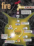
Fuel for the Fire
|
||||||||
“It struck me that NAD+ is strategically positioned — even uniquely positioned — to coordinate blood flow with energy metabolism.” JOSEPH R. WILLIAMSON, MD
“What we wanted to do is make sure this phenomenon is actually relevant to human work. There are many ways of monitoring signals in the animal brain, but we don’t have that many choices in doing human experiments. And so we have learned to depend on this increased blood flow signal. It then becomes very important to understand exactly what that signal represents.” MARK A. MINTUN, MD
The new discovery is expected to generate a rush of interest due to its potential impact on scientists around the world using changes in blood flow to map human brain function.
|
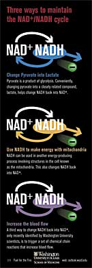
A CENTURY-OLD MYSTERY is taking place in your head as you read these words. Blood vessels in regions of your brain are widening, bathing cells in an increased blood flow. Scientists have known for more than 100 years that these changes take place when areas of the brain become activated or when any cell, such as a muscle involved in an exercise routine, increases its workload. They once assumed that the change occurs to supply cells with more of the glucose and oxygen that they needed to fuel their increased workload. Thanks in large part to researchers at Washington University in St. Louis, though, that old explanation has fallen away. Left in its place is a puzzle: If increased blood flow isn’t needed to supply cells with more fuel, then what exactly is it providing? Researchers Joseph Williamson and Mark Mintun don’t have the full answer yet, but with a pair of papers published early this year in the Proceedings of the National Academy of Sciences (PNAS), they moved the scientific community a major step closer to it. Williamson and Mintun have found the answer to a closely related question: How are blood flow increases triggered? Study results reported in PNAS link the increases to a molecule that occupies a unique and central spot in cellular energy production. The investigators hope to apply the new insights to improve imaging of the brain in action and to limit the side effects of diabetes, but their findings also are likely to have ramifications that ripple out far beyond their research specialties. Knowing how increased blood flow in the brain is activated could be relevant, for example, to understanding and controlling Alzheimer’s disease and stroke. Washington University researchers have been leaders in the development of functional brain imaging techniques, many of which monitor changes in brain blood flow levels. They first began to topple the old explanation for increased blood flow in 1988 by looking more closely at what brain blood flow changes reflected. “Much to our great surprise, what we observed was that when blood flow did come up in an active area of the brain, the amount of oxygen being used didn’t,” says Marcus E. Raichle, MD, professor of radiology, neurology and of anatomy and neurobiology. Raichle and other School of Medicine researchers confirmed and expanded the findings over several years, showing that brain activation increased blood flow but produced only a moderate increase in sugar use and a very small increase in oxygen use. “What remained was still the question of how are blood flow increases orchestrated and why?” Raichle recalls. The answers remained dauntingly out of reach until Washington University pathologist Joseph R. Williamson, MD, now retired, happened onto the search in the mid-1990s. With the support of the St. Louis-based Kilo Diabetes and Vascular Research Foundation and the National Institutes of Health, Williamson was studying the damaging effects of diabetes, which, in addition to elevating sugar levels, increases blood flow and harms blood vessels in the nerves, heart, retina and kidneys. Wondering if connections might exist between the increases in blood flow brought on by brain activity and those triggered by diabetes, Williamson found Raichle’s 1988 study and read it. As he investigated the scientific record further, Williamson recognized a similarity between working muscle cells and endangered cells in people with diabetes: both experienced increases in the ratio of two forms of a compound in energy metabolism, nicotinamide adenine dinucleotide (NAD). “It struck me that NAD is strategically positioned — even uniquely positioned—to coordinate blood flow with energy metabolism,” says Williamson. Biochemists who study cellular energy production put NAD at the center of a complex flow chart linking two different methods of producing the energy that powers most cells. For these methods, NAD serves as the major carrier of protons and electrons. Most NAD in the body is in an oxidized form scientists refer to as NAD+. During one method, glycolysis, a process that rapidly produces energy from sugar, electrons and protons are transferred from sugar to NAD+, changing it to NADH (NAD+ plus a proton and two electrons). “Not only is glycolysis twice as fast, it doesn’t require oxygen,” Williamson notes. “It’s really vital for survival.”
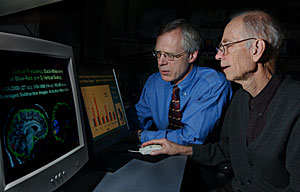
Mark A. Mintun, MD, and retired pathologist Joseph R. Williamson, MD, are studying blood flow in the brains of both rats and humans. Rapid glycolysis depends on a low ratio of NADH/NAD+. In activated cells and in cells endangered by diabetes, molecules of NADH increase, driving the NADH/NAD+ ratio up. Williamson suspected the ratio might be controlling changes in blood flow. He thought he could test his theory using a 50-year-old
link between the NADH/NAD+ ratio and the ratio of two other compounds
involved in energy production, lactate and pyruvate. This link was the
tool he needed to be able to alter NADH/NAD+: inject lactate or pyruvate,
change the NADH/NAD+ ratio, and see if blood flow increased or decreased. In a recent paper, published in January in PNAS, Williamson’s group confirmed the link again in studies of the rat retina and the visual region of the rat brain. They also identified a signaling pathway in cells that is triggered by high NADH/NAD+ ratios. The pathway activates a chain reaction that recycles NADH back into NAD+ and also promotes the production of nitric oxide, which dilates blood vessels. Williamson shared the successful results of the second rat experiments with Raichle in advance of publication, and they made the idea of testing the same principles in humans irresistible to Mark A. Mintun, MD, professor of radiology and of psychiatry. “What we wanted to do is make sure this phenomenon
is actually relevant to human work,” Mintun says. “There are
many ways of monitoring signals in the animal brain, but we don’t
have that many choices in doing human experiments. And so we have learned
to depend on this increased blood flow signal. It then becomes very important
to understand exactly what that signal represents.” Vlassenko is clearly pleased with the results: without lactate injections, the blood flow increase to the visual cortex during the visual task was 19 percent; after lactate injections, it was 26 percent. “That might not seem like a lot if you look strictly at the gain, but if you look at the gain as a percentage of original level of increase, that’s fully one-third more,” Vlassenko says. Follow-up work to the human study is generating “gorgeous”-looking data, according to Mintun. He expects the new discovery to generate a rush of interest due to its potential impact on scientists around the world using changes in blood flow to map human brain function. Williamson notes that while he’s not going to be conducting any follow-up experiments per se, he does plan to use the studies of the rat retina and data from other pre-retirement experiments to advance a new theory he has about how diabetes damages tissues. He thinks the culprit may be increased metabolism of glucose to fructose, which also increases the ratio of NADH/NAD+. Convincing colleagues in diabetes research to take a closer look at the mechanism has been an uphill battle; however, he is optimistic that as investigators understand that the NADH/NAD+ ratio regulates blood vessel function as well as energy metabolism, they will take a closer look at its role in damaging blood vessels and nerves in patients with diabetes. “There’s enough recent information now supporting
the importance of this mechanism that I think more people will be convinced
of its significance in the near future,” he says.
|
|||||||