
|
|
||
|
Nanomedicine Microscopic particles aid diagnosis and treatment of disease
|
|||||||
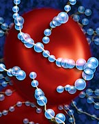
Nanoparticles have an inert core coated with a layer of active components — homing agents, imaging substances or drugs. Here, the particles (blue) are engineered to target strings of fibrin in a blood clot.
Nanoparticles have great potential for spotting disease
sites at the early stage when treatment is most effective. They also
can take uncertainty out of patient follow-up
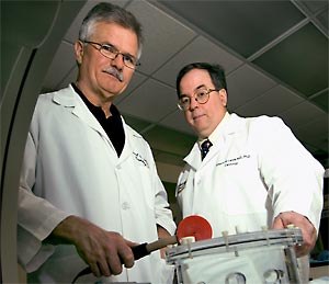
| IMAGINE replacing numerous medical tests, scans or surgeries with a simple injection. That’s the hope offered by nanoparticles — extremely small, bead-shaped carriers of medicinal agents. Using these tiny spheres, doctors will be able to locate disease sites deep within the body and at the same time determine their size, shape and diagnostic features. They can then adapt the nanoparticles to deliver a load of therapeutic drugs exactly where needed. Readily manufactured to contain a variety of components, the particles can be tailored to each patient’s unique condition. Co-inventors Samuel A. Wickline, MD, and Gregory M. Lanza, MD, PhD, have already seen their technology work against such major health challenges as cancer and cardiovascular disease in laboratory animals. Now they are set to test its effectiveness in human patients.
Samuel A. Wickline, MD, and Gregory M. Lanza, MD, PhD homing in and lighting up Although the nanoparticles are a few thousand times smaller than the dot above an “i,” each can carry hundreds of thousands of molecules on its surface. To induce the particles to home in on a target, the researchers attach molecules on the surface that recognize and bind to complementary molecules on the surface of target cells, whether the target is a cancerous tumor or a plaque in an artery. A suspension of the nanoparticles can then be injected into the bloodstream where the particles travel until they reach their objective. The ability to target internal disease sites with an externally administered therapy is a leap forward, but the ability to visually confirm that they are reached adds an important dimension. When loaded with an imaging agent in addition to a homing agent, the particles can both find disease sites and “light them up”; that is, make them easy to visualize using widely available scanning methods such as MRI, CT or ultrasound. “ In the past, experiments with targeted molecular imaging using other types of carriers failed because the signal at the target site was too faint,” says Lanza, associate professor of medicine and a cardiologist at Barnes-Jewish Hospital. “But our nanoparticles vastly increase the density of imaging agent at the site so that the signal becomes quite strong.” That’s because the amount of surface area available for attaching imaging agents to the nanoparticles is enormous. Say you have a cube just one inch on each side. To cover that cube would require just six square inches of paint, a spot about two and one-half inches wide. But if you cut the cube into a bunch of tiny cubes about the size of the nanoparticles, you would need 600,000 square inches of paint to cover them. That’s enough paint for a wall 10 feet high and 400 feet long. In a set of laboratory experiments, the researchers loaded their particles with about 100,000 molecules each of a metal that provides contrast in MRI scans and a few thousand molecules of an agent that homed in on growing tumors. The high payload of metal molecules allowed the researchers to see tiny, two-millimeter-wide melanoma tumors in mice using MRI scans identical to those in standard use for human heart or brain scans. The same tumors were invisible when scanned in the absence of the nanoparticles. Because they can pinpoint and then illuminate tiny regions, the nanoparticles have great potential for spotting disease sites at the early stage when treatment is most effective. They also can take uncertainty out of patient follow-up by definitively highlighting areas where disease symptoms have just begun to recur.
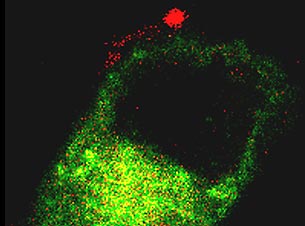
Delivered via nanoparticle carriers, drug components and lipids enter the membrane of a targeted melanoma cell. targeted treatment With the capacity to carry therapeutic agents also in the nanoparticles’ array, doctors will have a full-function medical missile available to them. “We’ve reached the era of targeted drug delivery where you now can see drugs being delivered to the right place,” asserts Wickline, professor of medicine and also a cardiologist at Barnes-Jewish Hospital. “ With nanotechnology, we’ve been able to bring Paul Ehrlich’s concept of a magic bullet to realization,” Lanza adds. Ehrlich, winner of the 1908 Nobel Prize in Physiology or Medicine, sought compounds that would be specifically attracted to and kill disease-causing organisms, with no harmful effect on the body. “ Like Ehrlich’s magic bullet, our nanoparticles are attracted to a particular disease site. Because they accumulate at a well-defined location, they have little effect on the rest of the body,” Lanza says. In contrast, standard drug administration dilutes medication throughout a person’s system. While a portion of the medication eventually reaches the area where it is needed, it also goes where it isn’t needed in equally toxic concentrations. Already-tested versions of the nanoparticles locate tumors and atherosclerotic plaques using small homing molecules attracted to proteins abundant on the cells of newly forming blood vessels. As tumors grow, they recruit new blood vessels to obtain nutrition. Arterial plaques also stimulate the growth of capillaries around them. So, both abnormal growths can be detected using the same homing strategy. Drugs specifically designed to kill cancer cells or to dissolve plaques can be included on these nanoparticles along with homing and imaging agents. Then the brightness of the image at the site reveals both the size of the lesion and the amount of drug that reached the site. The treating physician would have complete information about dose achieved the same day the treatment was given and could quickly adjust treatment as needed. personalization “ The nanoparticle technology is really the next step in personalized medicine,” Wickline says. “There’s a lot of interest in individualizing medicine by analyzing each patient’s genome. The genome interacts with its environment in very complex ways so that the results often can’t be predicted exactly.” “ On the other hand,” Lanza says, “the nanoparticles locate what’s already present in the body.” “ Right,” Wickline says. “We’re not reading the script — the genes — we’re looking at the actors — the proteins. We’re the critics watching the play as it happens, not reading it as it was written.” That means that each patient’s health status can
be analyzed and treated as a unique case, instead of a generalization.
The nanotechnology
can be designed “ Our nanotechnology is very flexible,” Wickline says. “During the course of treatment, a doctor could decide to target a different biomarker or numerous markers simultaneously.” Flexibility allows the nanoparticles to be loaded with combinations of compounds and used as a one-step diagnostic test. One injection could reveal the proportions of different biomarkers and indicate the nature of a disease — how aggressive a cancer is or how likely a plaque is to rupture, for example. The medical school’s BioMed 21 initiative calls for efficient development of laboratory discoveries into practical medical applications, and Wickline and Lanza have support from both the medical school and corporate partners to ensure their technology will be rapidly put to use for the benefit of patients. They have built collaborations with Phillips Medical Systems, Dow Chemical, Bristol-Meyers Squibb Medical Imaging, and a local company they founded called Kereos Inc., which will set up clinical trials. They also have received $15 million in support from the National Institutes of Health. “ We’ve maintained control of the technology instead of licensing it out so that we can make sure it develops to its fullest potential,” Lanza says. “
This technology can strongly affect the practice of medicine, and we intend
to have it do so,” Wickline emphasizes.
|
||||||