


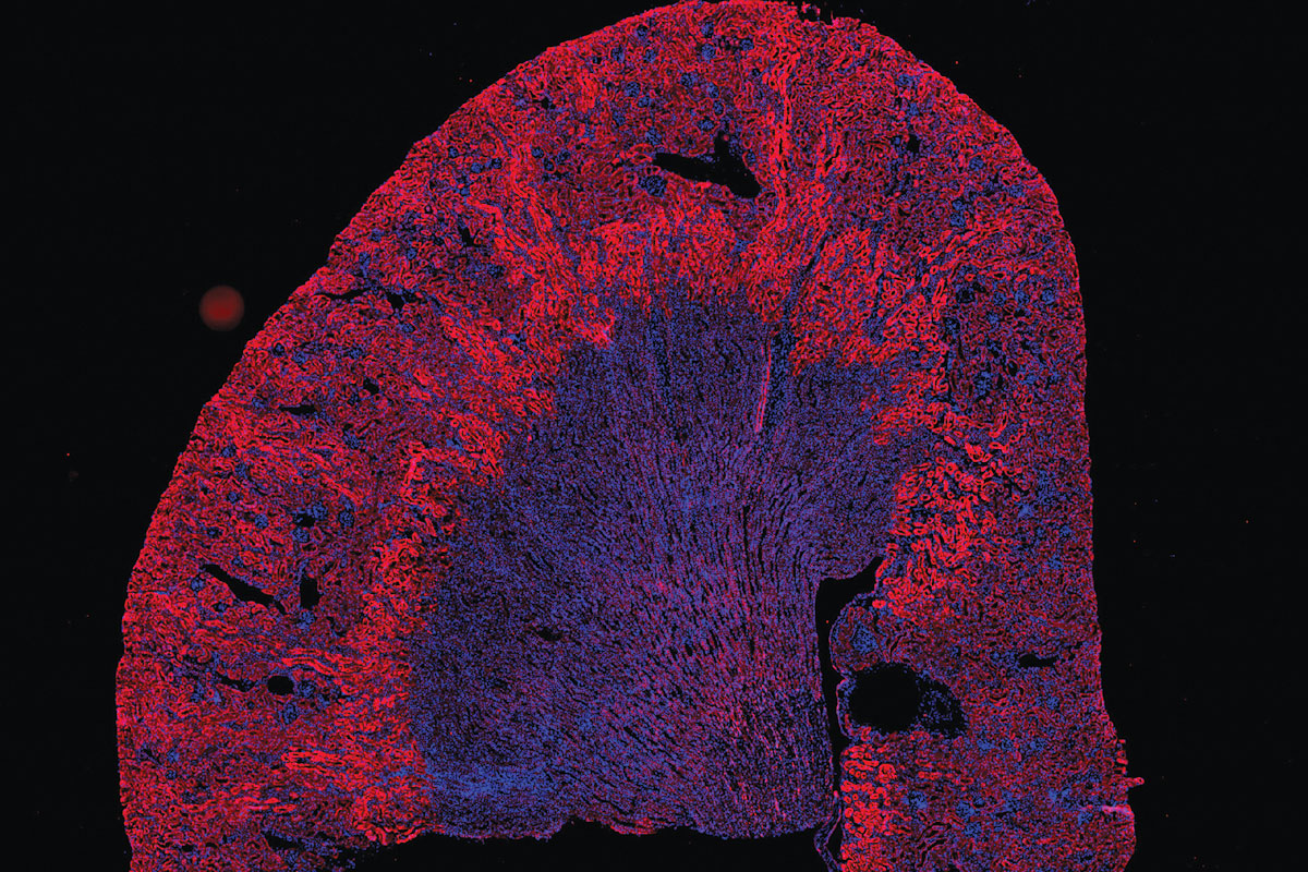
Cells near the edge of a mouse kidney glow red, a sign that they are starved for oxygen. This is an important clue for researchers studying how the kidney responds to injury. Image taken on Zeiss Axioscan Z1 Automated Slide Scanning System.
Twelve feet tall and weighing several tons, the microscope capable of generating the voltage of a stun gun looms over James Fitzpatrick, PhD, as he circles it, deftly opening latches and detaching panels. A door on the side swings open, revealing a tangle of wires and, near the top, a shelf the size of a tape cassette. When the machine is fully built and turned on, a robotic arm will reach down, pick up experimental samples placed on the shelf and insert them into a column deep inside. There, the samples will be bombarded with electrons.
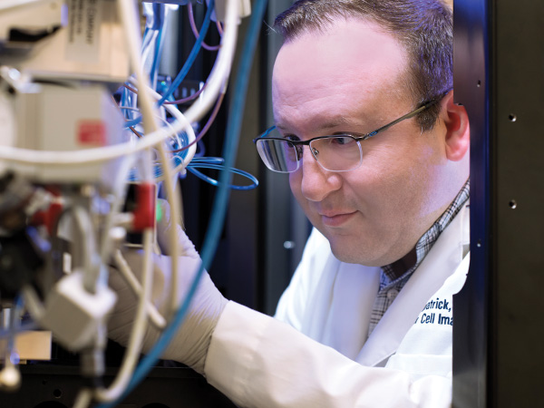
James Fitzpatrick, PhD, adjusts the wiring inside the center’s new cryo-electron microscope. The instrument visualizes objects as small as a few billionths of a meter, allowing researchers to map the location of each atom in a protein.
And the invisible will be unveiled.
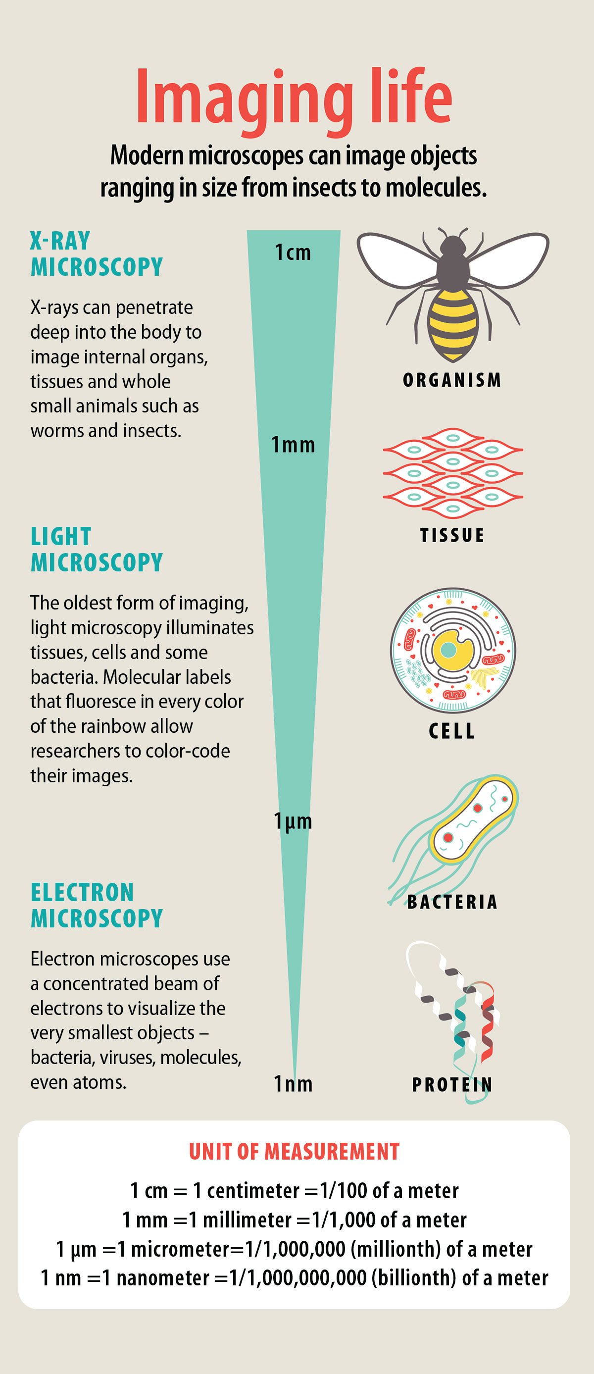
This instrument that Fitzpatrick has so painstakingly unsealed is a top-of-the-line cryo-electron microscope. It is capable of magnifying the living world’s tiniest structures — bacteria, viruses and biomolecules — a million or more times, bringing the unimaginably small up to a human scale.
“It’s a complicated beastie,” said Fitzpatrick, grinning. He is director of the Washington University Center for Cellular Imaging, known as WUCCI and pronounced Wookiee. He has spent the better part of the spring and summer overseeing the installation and calibration of the $7.2 million microscope, and he will put it through weeks of testing before making it available to the center’s clients later this fall. The machine is the cornerstone of a significant investment in cellular imaging. Advances in the field are transforming biomedical research, and WUCCI is keeping Washington University at the forefront.
Introducing the WUCCI
WUCCI sprawls across 6,000 square feet in the basement of the McKinley Research Building (to be renamed the Debra and George W. Couch III Biomedical Research Building) on the Medical Campus. It houses 15 of the most advanced microscopes, as well as banks of high-powered computers that are needed to analyze the mountains of data they produce.
The scopes can take 3-D images of viruses, make movies of living neurons as they signal one another, visualize the beating hearts of small animals such as worms and fish and map the structure of proteins down to the location of each atom. Since the center’s establishment in 2015, researchers have imaged everything from living zebrafish and butterfly eyes to plastic catheters and rocks.
Today, more than 500 Washington University researchers representing some 220 labs use the center, and Fitzpatrick is hoping to draw more.
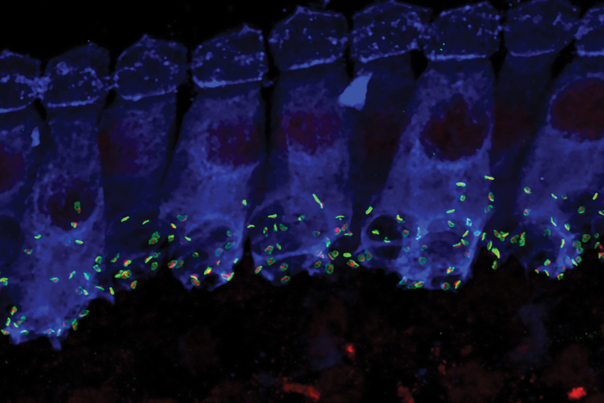
To study how sound is transmitted from the ear to the brain, researchers stained specialized sound-detecting cells (blue) and their synapses (red/green) in a mouse inner ear. Image taken on Zeiss LSM 880 Airyscan Microscope.
Cornerstone of biology
When Anton van Leeuwenhoek looked through the first microscope more than 300 years ago and saw legions of previously unimagined life forms wriggling underneath, he established one of the foundations of modern biology.
“Imaging is one of the cornerstones of biology and biomedical science,” said Azad Bonni, MD, PhD, the Edison Professor and chair of the Department of Neuroscience. “Whatever questions we’re trying to answer, it all comes down to cells, because they are the fundamental unit of living things.”
Modern research microscopes are as far from Leeuwenhoek’s single-mounted lens as smart phones are from Alexander Graham Bell’s invention. They not only take snapshots of cells mounted on slides, they can record movies of a fertilized egg developing into an embryo, or of an immune cell chasing a bacterium. They can focus deep inside bones, tumors and even small animals, allowing scientists to see cells and tissues in their natural environment. They reveal our DNA. Microscopy makes the invisible visible.
WUCCI builds on the strengths of the departments of Neuroscience and of Cell Biology and Physiology, which operated separate imaging facilities for a number of decades. About five years ago, at funding renewal time, department leaders questioned whether to re-establish separate facilities or join together. “It occurred to us that it might be a good idea to create a collaborative center greater than the sum of its parts,” Bonni said.
X-ray tomography detects an emulsion of gold nanoparticles, tracing the network of blood vessels in a mouse’s lungs and heart. Image taken on Zeiss Versa 520 X-Ray Microscope.
Credit: Matthew Joens (WUCCI) and Kel Vin Woo (Ornitz Lab)
The neuroscience imaging facility specialized in light microscopy, which allows researchers to attach fluorescent markers to specific molecules or cells, color-coding their images. Such technology suits researchers who often study how cells respond to cues, and circuits of cells and the synapses connecting them.
Cell biologists lean more heavily on electron microscopy, which images objects tens to thousands of times smaller than cells. Electron microscopes reveal precisely how the essential molecular machinery of the cell operates and how molecules involved in disease might be targeted with drugs.
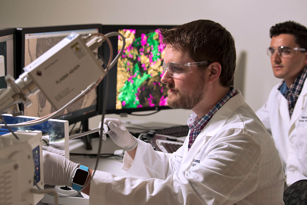
Staff scientist Matt Joens, left, and research assistant Daniel Geanon.
Members of both departments — Paul Taghert, PhD, and Bonni from Neuroscience, and Helen Piwnica-Worms, PhD, Robert Mecham, PhD, David Piston, PhD, and Phyllis Hanson, MD, PhD, of Cell Biology and Physiology — advocated for this core center and engaged in discussions across the medical school. Taghert and Hanson traveled the country, gathering the best ideas from leading imaging facilities. Ultimately, Fitzpatrick was recruited from The Salk Institute in San Diego.
“We sold him on the idea that he could build something big here,” Bonni said.
Institutional resource
Modern imaging is key to understanding and treating many diseases. Thirty years ago, there were no microscopes that could map the blood vessels inside a solid tumor, or trace the outlines of each molecule in a virus’s shell. That technology now hums and clicks and reshapes medicine in Fitzpatrick’s center, and researchers increasingly rely on it. With every technological improvement, microscopy has grown more central to biological research … and more expensive.
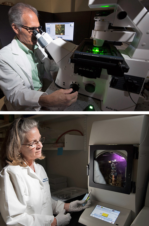
Research specialist Dennis Oakley, top, and instructor Robyn Roth.
“Many researchers need access to a wide range of imaging tools but could never afford to have them in their lab,” said Piston, the Edward Mallinckrodt Jr. Professor and head of Cell Biology and Physiology. “Often that means that young investigators have to make compromises on their choice of projects, but having access to WUCCI allows them to think more broadly.”
Furthermore, effectively using the high-tech equipment requires skills that can be difficult for small research groups to obtain.
“As the technology becomes more complicated, it takes more and more specialized expertise to get the most out of it,” Piston said. “In a large central facility, we can afford to hire and train the right specialists.”
Fitzpatrick likes to call the facility a one-stop shop, because the seven full-time staff members — all scientists themselves — are prepared to help with any stage of research.
A 3-D rendering of a pair of cancerous mouse kidneys rotates in the video above. The large, fluffy yellow bodies are tumor metastases, and the smaller yellow spots and squiggles are blood vessels inside the kidneys. The organs were imaged using an X-ray microscope that sends beams of X-rays slicing through the tissue to create hundreds of 2-D images, which are then digitally recombined into one 3-D visualization. The X-rays do not damage the tissue, allowing the tissues to be preserved for further study after imaging.
Credit: Michael Ross, Weilbaecher Lab and Matthew Joens, James Fitzpatrick, Center for Cellular Imaging
“I have sat down with people at WUCCI and talked about basic cell biology,” said Clifford Luke, PhD, an associate professor of pediatrics who has used the center’s super-resolution light microscopes. “If you tell them what question you’re trying to answer, they will help you figure out the best way to do the experiment and which microscope is best suited for it.”
WUCCI has a suite devoted to sample preparation, containing equipment such as diamond knives sharp enough to cut extremely thin slices of tissue. Just as sets for TV shows became more realistic as high-definition TVs became commonplace, sample prep has become more important — and more elaborate — as microscopy has improved.
“When the resolution wasn’t as good, the quality of the sample prep didn’t matter as much,” Fitzpatrick said. “If something was a bit distorted, you couldn’t tell. These days, the quality of sample prep is of paramount importance.”
Immense computing power is required to process the complex datasets that come off the microscopes. Powerful computers line one wall of the center, and center staff can assist with analysis.
Pushing the limits
WUCCI is the latest step in a long history of imaging strength and innovation at Washington University. The first electron microscope built in the U.S. was designed and constructed by Gordon H. Scott, PhD, associate professor of cytology in the Department of Anatomy, around 1935.
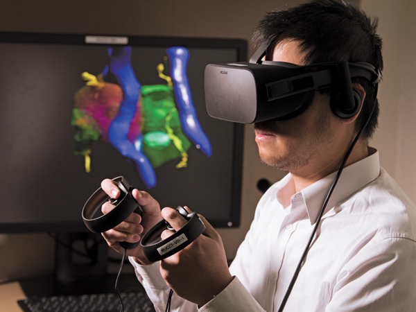
Wearing a headset and hand controllers, Michael Shih, PhD, explores the virtual reality scene he has created from a researcher’s 3-D still image.
More recently, Timothy Holy, PhD, the Alan A. and Edith L. Wolff Professor of Neuroscience, developed a form of light-sheet microscopy, which allows scientists to view thousands of cells at a time. Holy used this variation on the technique to scan more than 10,000 cells in a mouse’s nose to find the rare ones that turn on when the animal sniffs a particular pheromone.
Now, WUCCI staff are pushing the next frontier of imaging: virtual reality.
“We have microscopes that produce these beautiful, detailed 3-D images, but then we display them on a flat 2-D screen,” Fitzpatrick said. “We think that if people can take their results, hold them in their hands, turn them around, and look at them from all angles it will generate new ideas and understanding.”
Michael Shih, PhD, a center research specialist, has begun developing software that can convert researchers’ 3-D images into a virtual reality. The goal is to create an immersive environment to navigate biological samples ranging in scale from molecules to organ systems.
Although a virtual still image allows researchers to manipulate and inspect a virtual object, a virtual reality movie would allow people to watch from the inside as a biological process took place. Imagine standing at a cell’s midline and watching as the chromosomes lined up around you and then pulled apart into two daughter cells.
“Hardly anyone is working on the applications of virtual reality in microscopy,” Fitzpatrick said. “This is something we are working on offering that no other imaging facility can do.”
Looking ahead
WUCCI has the potential to transform how biology is done at Washington University. The center ranks among the top five best in the U.S., as measured by the quality of the microscopes and the power of the computers. Staying in that position requires an ongoing commitment. Increasingly, other departments are contributing specialized equipment. The cryo-electron microscope, for instance, was jointly purchased by the offices of the provost and the medical school dean and other medical school departments led by Biochemistry & Molecular Biophysics and Cell Biology & Physiology.
“More and more, biomedical research depends on new technology,” Piston said. “There has been a lot of expertise in imaging at Washington University for a long time, but it has been distributed in different departments and centers, and it was difficult for researchers to find all the parts they might need, or to even learn that those were available. “With WUCCI, we now have the ability to access the newest technology through shared contributions.”

 Share
Share Tweet
Tweet Email
EmailPublished in the Autumn 2017 issue